
Jun
If you click on links we provide, we may receive compensation.
The epidermis is only about 0.1 millimetres thick on your eyelids – roughly the width of a human hair – yet inside that sliver of tissue, at least four specialised cell types are working simultaneously to manage UV protection, immune surveillance, hydration control, pigmentation, and the continuous renewal of your skin’s outer surface. Research has found that epidermal cell turnover slows by up to 50 percent in older adults compared to younger skin, and that slowdown is behind more of what we call “aging skin” than most people realise. (Source: Cellular Senescence in Human Skin Aging, PMC, https://pmc.ncbi.nlm.nih.gov/articles/PMC10873061/) Getting to know the epidermis is not an exercise in biology for its own sake. It is the fastest route to understanding why some skincare ingredients actually deliver results – and why some others, regardless of what the label says, never reach their target.
This is the second piece in our foundational skincare science series. The first covered the stratum corneum in detail – that outermost cellular layer where the skin’s famed “brick and mortar” barrier lives. This article zooms out one level to look at the full epidermis: what it is, what it contains, how it changes with age, and what a well-chosen skincare routine can realistically do to it. Consider this the map to the territory. Once you have it, ingredient lists start to make a lot more sense.
What Exactly Is the Epidermis?
Your skin has three main layers. The epidermis is the outermost – the part you see and touch, the skin’s primary interface with the outside world. Beneath it sits the dermis, which contains collagen, elastin, blood vessels, and most of the structural architecture responsible for skin’s firmness and resilience. Below that is the hypodermis, the fatty subcutaneous layer that insulates and cushions. The epidermis is where everything visible happens. It is also the layer that skincare products interact with directly.
Its thickness varies considerably depending on where you are on the body. On the eyelids, the epidermis measures roughly 0.1 millimetres – thinner than a sheet of paper. On the palms of the hands and the soles of the feet, which bear constant friction and pressure, it can reach 1.5 millimetres or more. On the face – where most of us focus most of our skincare attention – it typically falls between 0.05 and 0.1 millimetres. That is not a lot of material to protect the rest of you. It is also not a lot of material to recover from damage, which has direct implications for how aggressively we can treat it.
One of the most striking facts about the epidermis is what it does not have: blood vessels. Unlike almost every other tissue in the body, the epidermis receives no direct blood supply. Every nutrient, every oxygen molecule, every immune cell that reaches it must diffuse upward from the dermis below, crossing the epidermal-dermal junction and making its way through the living layers of the epidermis from the inside out. That dependence on diffusion makes the health of the junction between the two layers critically important – and, as we will see, it is one of the things most significantly altered by aging.
What Are the Four Layers of the Epidermis?
The epidermis is divided into four distinct layers in most of the body (a fifth layer, the stratum lucidum, appears only on the thick skin of the palms and soles). Each layer represents a stage in the life of a skin cell – a journey that begins at the bottom and ends at the surface. Understanding what happens in each layer makes it much easier to see where specific skincare ingredients intervene.
The stratum basale is the deepest layer, sitting right against the epidermal-dermal junction. This is where new keratinocytes are born. Stem cells here divide continuously, pushing daughter cells upward to begin their differentiation journey. The stratum basale also contains melanocytes – the pigment-producing cells – as well as Merkel cells, which are involved in sensory touch detection. And this is where vitamin D synthesis begins: UVB radiation interacting with 7-dehydrocholesterol stored in basal keratinocytes converts it into previtamin D3, which then isomerises into active vitamin D3. Research confirmed by the Linus Pauling Institute at Oregon State University identifies the stratum basale and stratum spinosum as the layers carrying the highest concentrations of this precursor compound. (Source: Vitamin D and Skin Health, Linus Pauling Institute, https://lpi.oregonstate.edu/mic/health-disease/skin-health/vitamin-D)
Above the basale sits the stratum spinosum – the spiny layer, so named because the cells’ surfaces bristle with tiny protein projections that lock neighbouring cells together through structures called desmosomes. Keratin production is well underway here. Small organelles called lamellar bodies begin forming in spinosum cells; these are loaded with lipids and enzymes that will eventually be secreted higher up to form the waterproof matrix of the stratum corneum. The immune cells known as Langerhans cells live primarily in this layer, sampling the chemical environment around them for anything that should not be there.
The stratum granulosum follows, named for the dark protein granules visible inside the cells – keratohyalin granules, which help pack keratin fibres tightly as the cell prepares to die. This is also where lamellar bodies release their lipid contents into the intercellular spaces between cells – the beginning of the lipid bilayers that define the stratum corneum’s barrier function. Cells in the granulosum are flattening, losing their nuclei, and completing the transformation from living keratinocyte to corneocyte. And finally, the stratum corneum – the outermost layer, the subject of our companion piece – is where the cells arrive at their destination: completely flat, metabolically dead, embedded in layers of ceramides, fatty acids, and cholesterol, forming the physical and chemical barrier that protects everything underneath.
Advertisement
Who Actually Lives in the Epidermis?

Keratinocytes make up 90 to 95 percent of the epidermal population. They are the workhorses: producing keratin for structural strength, forming tight junctions to make the barrier selective, and generating the lipids that cement the stratum corneum together. In young adults, the full journey from stratum basale to stratum corneum takes roughly 28 to 45 days. That timeline is what people refer to when they talk about skin cell turnover. It is the underlying rhythm of the skin’s self-renewal – and retinoids, among other things, work partly by accelerating it. With age, that rhythm slows noticeably, with consequences for texture, tone, and the skin’s ability to repair itself after injury.
Melanocytes make up about 5 to 10 percent of basal layer cells. They sit in the stratum basale and produce melanin – the pigment responsible for skin colour, freckles, and the darkening response to UV exposure. Melanin is synthesised through a process called melanogenesis, packaged into organelles called melanosomes, and then transferred to surrounding keratinocytes. Research from PNAS has documented how keratinocytes actively regulate this transfer process via extracellular vesicle signalling, with the receiving keratinocyte playing a more active role than was previously understood. (Source: PNAS 2024, https://www.pnas.org/doi/10.1073/pnas.2321323121) Once inside the keratinocyte, the melanin positions itself directly over the cell’s nucleus – sitting there like a tiny umbrella, absorbing and scattering UV radiation before it can damage the DNA below. This dual role as both pigment and shield is why melanin is simultaneously a cosmetic concern and a protective mechanism, and why decisions about pigmentation treatments are never entirely separate from UV protection.
Langerhans cells are the epidermis’s immune officers. Derived from bone marrow precursors and belonging to the dendritic cell family, they sit mainly in the stratum spinosum and continuously sample the chemical and molecular environment around them. When they detect a potential threat – an allergen, a sensitiser, a hapten from a fragrance or preservative – they pick it up, migrate to regional lymph nodes, and present it to T cells to trigger an adaptive immune response. Research published in the Journal of Clinical Investigation has established that Langerhans cells also play a tolerance-setting role, educating the immune system to distinguish between genuinely dangerous exposures and harmless repeated contacts. (Source: JCI, https://www.jci.org/articles/view/63190) That dual function – recognising threats while also establishing tolerance – is part of the reason skin reactivity can shift in unpredictable directions, particularly as the Langerhans cell population changes with age.
Merkel cells round out the epidermal cell population, found in the stratum basale in association with nerve endings. They function as sensory receptors for light touch and texture discrimination. They are not a primary target for skincare, but their presence in the basale layer is a reminder that the epidermis is not just a barrier – it is a sensing surface, continuously gathering information about the environment and relaying it inward.
What Does the Epidermis Actually Do?
The full function list is longer than most people expect. The epidermis manages physical barrier integrity, UV protection and pigmentation, immune surveillance, hydration regulation, and vitamin D synthesis – simultaneously, in a structure that is often less than a tenth of a millimetre thick. Each of these functions depends on the continuous, coordinated activity of the cell types described above. When any part of that system is disrupted – by age, UV damage, harsh products, or inflammatory skin conditions – the downstream effects touch all the others.
The physical barrier aspect is the most discussed. The epidermis as a whole is what stands between the body’s interior and the external world. The stratum corneum does the most specialised barrier work – controlling water loss, resisting chemical assault, managing surface pH – but the viable layers beneath it are what continuously produce and refresh that outer defence. Think of the stratum corneum as the finished wall and the rest of the epidermis as the factory that keeps building it. If the factory slows down or the construction materials are depleted, the wall starts to thin.
Hydration regulation is worth separating from the simple idea of moisturisation. The epidermis does not store water like a reservoir. What it controls is transepidermal water loss – the rate at which water evaporates from the skin’s surface. When the epidermal barrier is intact and the lipid matrix is healthy, that loss stays minimal. When it is disrupted – by aggressive cleansing, over-exfoliation, or inflammatory conditions – water escapes faster and skin feels tight, dry, and reactive. The stratum corneum does the heavy lifting here, but the whole epidermal system is what allows the stratum corneum to exist in the first place.
Immune surveillance through Langerhans cells means the epidermis is, among other things, a first-line security checkpoint. Every product applied to skin, every ingredient in every formulation, passes the scrutiny of Langerhans cells. Most things are cleared without incident. But this is why some ingredients produce sensitisation reactions that worsen with repeated exposure, and why the immune health of the epidermis has real practical implications for how skin tolerates the routine you put it through.
How Does the Epidermis Change as We Age?

Age changes the epidermis in ways that are gradual and interconnected. None of them happen overnight. But together, they shift the way skin looks, feels, behaves, and responds to products in ways that are distinctly different from younger skin – and that understanding should shape every product choice.
Keratinocyte turnover slows. Research has found that epidermal cell turnover can be reduced by up to 50 percent in older adults compared to younger skin. (Source: Cellular Senescence in Human Skin Aging, PMC, https://pmc.ncbi.nlm.nih.gov/articles/PMC10873061/) What this means visually is dullness – older corneocytes stay at the surface longer, and the skin can look flat and uneven rather than fresh and light-reflective. It also means wound healing slows considerably. A barrier breach that younger skin might close in a few days can take the better part of two weeks in older skin. Corneocytes also become larger as turnover slows, which contributes to rougher texture and less consistent light diffusion across the surface.
Melanocytes decline in overall number – roughly 10 to 20 percent per decade after early adulthood – but the ones that remain tend to behave less consistently. Rather than an even distribution of active pigment-producing cells, what older skin has is a patchier arrangement: some areas underpigmented, some areas where clusters of melanocytes produce more melanin than neighbouring areas. The visible result is age spots, uneven tone, and a loss of the consistent translucency that characterises young skin. UV exposure accelerates this process considerably, which is why age spots appear primarily on chronically sun-exposed areas of the body.
Langerhans cell numbers fall with age, with some estimates suggesting a reduction of up to 50 percent in older adults. This has two related practical consequences. The first is reduced immune competence in the skin – older epidermis is genuinely less effective at mounting defence responses. The second is increased skin sensitivity: with fewer Langerhans cells to moderate immune responses, the threshold for irritation can shift. Ingredients that were tolerated for decades can suddenly become problematic. And the skin’s ability to establish new tolerance to exposures it has not encountered before may also diminish.
Epidermal thinning overall is another well-documented change. The epidermis becomes thinner, more transparent, and more fragile. Injuries that younger skin absorbs without visible consequence – friction from clothing, adhesive tape removal, a minor bump – can cause tearing or bruising in older skin. Basal keratinocytes themselves change shape, becoming shorter and wider rather than maintaining the tall, columnar form of younger cells, and this architectural change affects the structural integrity of the entire layer.
Perhaps the most clinically significant aging change is at the epidermal-dermal junction – the boundary between the epidermis and the dermis below. In young skin, this junction has a wavy, interlocking architecture formed by structures called rete ridges, which dramatically increase the surface area of contact between the two layers. More surface area means more efficient diffusion of oxygen and nutrients from the dermis into the epidermis. Research published in a detailed review of skin aging pathobiology has documented how this junction progressively flattens with age, reducing the interface area and impairing nutrient exchange. (Source: The Pathobiology of Skin Aging, ScienceDirect, https://www.sciencedirect.com/science/article/pii/S0002944020301425) The flattening also reduces the mechanical grip between layers – which is a primary reason mature skin bruises and tears more easily. Less anchoring means less resistance to shearing forces. And less nutrient exchange means the epidermis, which already has no blood vessels of its own, becomes progressively less well-supported from below.
What Can Skincare Actually Reach in the Epidermis?
The epidermis is selective about what it allows through. The stratum corneum exists partly to keep things out – pathogens, toxins, foreign molecules – which means penetration in skincare is not automatic and is never guaranteed just because a product contains an active ingredient. The concentration listed on a label, the quality of the ingredient, and the delivery system all determine whether what you apply actually reaches the target layer.
The key rule used by formulators and dermatologists is the 500 Dalton rule: molecules with a molecular weight below roughly 500 Daltons can penetrate the stratum corneum passively; larger molecules generally cannot. Retinol (approximately 286 Daltons), niacinamide (122 Daltons), and L-ascorbic acid, the most bioactive form of vitamin C (176 Daltons), all fall well within this range and can reach the viable epidermis. Collagen in a moisturiser – with molecular weights typically ranging into the hundreds of thousands of Daltons – cannot penetrate. It works at the surface, providing temporary hydration and a smoothing film effect, but it does not repair collagen from the outside in. That distinction matters when evaluating what a product can honestly be expected to do.
Vehicle matters enormously too. How an ingredient is delivered affects whether it reaches its target. Oil-soluble actives penetrate the stratum corneum’s lipid matrix more readily than water-soluble ones under ordinary conditions. Encapsulation technology – where an active is enclosed in a lipid or polymer shell designed to release at the target depth – can improve both penetration and stability for actives that might otherwise degrade before arriving. pH affects penetration in a more specific way: L-ascorbic acid needs a formulation pH below roughly 3.5 to penetrate effectively, because only the un-ionised form of the molecule crosses the lipid barrier easily. A vitamin C serum at pH 6, however elegantly packaged, delivers a fraction of the activity of one formulated at pH 3. (Source: Plastic Surgery Key, Percutaneous Delivery of Cosmetic Actives, https://plasticsurgerykey.com/9-percutaneous-delivery-of-cosmetic-actives-to-the-skin/)
How Do Key Ingredients Work at the Epidermal Level?
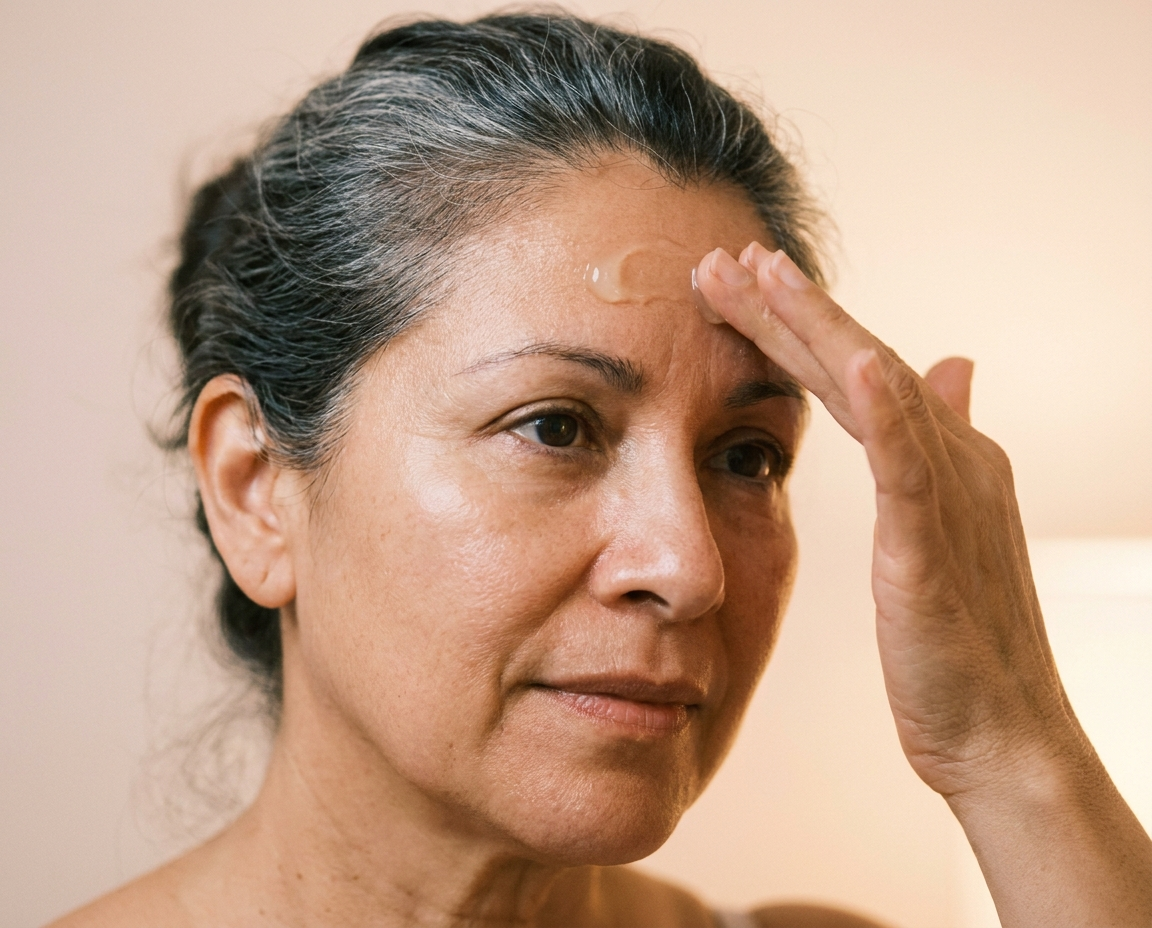
Retinoids are the most extensively studied topical skincare ingredient class, and their mechanism in the epidermis is well-established. Published research in PMC has shown that the retinoid signal is received by nuclear receptors in suprabasal keratinocytes, which then trigger a paracrine signalling cascade – involving heparin-binding EGF-like growth factor – that stimulates proliferation of basal keratinocytes from below. (Source: PMC, Physiological and retinoid-induced proliferations of epidermis basal keratinocytes, https://pmc.ncbi.nlm.nih.gov/articles/PMC125394/) The practical effect is accelerated keratinocyte turnover and, with consistent use, a thickening of the viable epidermis that counteracts age-related thinning. Retinoids also drive improvements in the epidermal-dermal junction architecture over time. These are not quick-fix effects – they accumulate over weeks and months of consistent use, and they reverse when use stops.
AHAs and BHAs – acids like glycolic, lactic, and salicylic – work primarily at the stratum corneum level, dissolving the protein bridges (corneodesmosomes) that hold dead skin cells to each other and to the surface. This accelerates desquamation – the shedding of corneocytes – producing the smoothing, brightening, and pore-clarifying effects that acid exfoliants are known for. The visible result is not the epidermis replacing itself faster from below; it is the epidermis revealing itself sooner by clearing cells that would otherwise linger on the surface. Consistent use also means the skin surface reflects light more evenly, and congested pores appear clearer as the accumulation of older cells is reduced.
Vitamin C works specifically at the melanocyte level, intervening in the melanin production pathway. L-ascorbic acid inhibits tyrosinase – the key enzyme in melanogenesis – interfering with the synthesis of melanin before it is produced and packaged. Research published in the International Journal of Cosmetic Science has documented this mechanism in detail. (Source: Melanogenesis and skin-lightening agents, IJCS, https://onlinelibrary.wiley.com/doi/10.1111/j.1468-2494.2010.00616.x) The result, with consistent use and adequate daily SPF, is a gradual lightening of areas where melanocytes have been overproducing. Because it targets the production step, vitamin C is most effective when paired with UV protection that limits the repeated UV triggers that drive melanocyte overactivity in the first place.
Niacinamide targets the same visible outcome – uneven pigmentation – but at a different point in the process. Rather than blocking melanin production, it inhibits the transfer of melanosomes from melanocytes to keratinocytes. A study cited in the British Journal of Dermatology found that niacinamide produced 35 to 68 percent inhibition of melanosome transfer in a co-culture model, with no effect on melanocyte activity itself – meaning melanin keeps being synthesised, but it is blocked from spreading through the skin. (Source: ResearchGate, Niacinamide and melanosome transfer, https://www.researchgate.net/publication/11276395_The_effect_of_niaciamide_on_reducing_cutaneous_pigmentation_and_suppression_of_melanosome_transfer) This different mechanism is why vitamin C and niacinamide complement each other well in brightening formulations – they address the same visible problem from opposite ends of the melanin pipeline.
SPF does considerably more for the epidermis than block sunburn. Daily sun protection reduces the repeated UV triggers that drive melanocyte overactivity and erratic pigmentation. It preserves Langerhans cell populations that would otherwise be depleted by UV exposure: studies in PubMed have shown that an SPF 30 sunscreen can substantially prevent UV-induced Langerhans cell depletion in skin. (Source: PubMed, Commercial sunscreen lotions prevent UV-induced depletion of Langerhans cells, https://pubmed.ncbi.nlm.nih.gov/9779495/) It limits the cumulative DNA damage in keratinocytes that accelerates structural aging. And it is one of a very small number of interventions supported by robust long-term evidence for actually slowing the visible aging of skin – not because it reverses existing damage, but because it reliably prevents new damage from accumulating.
Ceramides and barrier-support ingredients work at the stratum corneum surface rather than reaching the viable epidermis. But this still matters for the whole epidermal system. A compromised outer barrier triggers an inflammatory cascade – producing cytokines and other signalling molecules that can disrupt keratinocyte behaviour throughout the living layers of the epidermis. Keeping the barrier reinforced from the outside reduces the repair burden that falls on the basal layer, and it reduces the inflammatory environment that accelerates aging across the whole epidermal stack.
What Should Skin in Its 50s, 60s, and Beyond Know About All of This?

Mature skin is not damaged skin. It is skin operating on a different set of biological parameters – slower turnover, fewer melanocytes, a thinner epidermal layer, a flatter dermal junction, a reduced Langerhans cell population. Each of those changes is real and has practical consequences. But once you know what the parameters are, product choices become far less mysterious.
The slower keratinocyte turnover of mature skin means exfoliation matters more, not less, to maintain a clear, even surface. Without some regular help clearing older cells, skin can look dull and feel uneven. But the thinner, more fragile epidermis also means that aggressive exfoliation carries a higher cost. Daily high-concentration AHAs can compromise a barrier that is already working harder to maintain itself and has less structural reserve to recover from disruption. Most dermatologists recommend regular, moderate exfoliation for mature skin – two or three times per week with a gentle AHA or a well-tolerated enzyme exfoliant – rather than daily or high-strength treatments that strip more than they reveal.
Retinoids remain the gold standard in mature skincare, despite a persistent myth that older skin cannot tolerate them. The evidence base for retinoids in counteracting epidermal thinning, accelerating keratinocyte turnover, improving barrier integrity, and supporting collagen synthesis in the dermis is arguably stronger for mature skin than for any other age group – because the changes retinoids address are most pronounced in mature skin. The approach does shift with age: lower starting concentrations, less frequent initial application, and pairing with robust barrier support (ceramides, niacinamide, a simple but thorough moisturiser) makes a significant difference to tolerability. Well-formulated retinol in a barrier-supportive base is a very different experience from the same percentage applied to dry skin with no buffer.
The erratic melanocyte behaviour that comes with age makes consistent daily SPF more consequential, not less. Each unprotected UV exposure risks triggering a new cycle of melanocyte overactivity in areas where the cells are already producing pigment unevenly. An SPF of 30 or higher, applied every morning regardless of weather or season, is the single most effective thing someone in their 50s, 60s, or beyond can do to limit further pigmentation irregularity. Vitamin C and niacinamide work most effectively when SPF is already in place – without UV protection limiting the upstream trigger, the brightening work is ongoing rather than cumulative.
The flattening of the epidermal-dermal junction is not something topical products can fully reverse on their own. But supporting the living epidermis from outside – with barrier-appropriate ingredients, a retinoid used consistently, and rigorous UV protection – creates the best possible conditions for the junction to maintain its architecture as long as possible. And keeping the epidermis from being repeatedly disrupted reduces the inflammatory cycling that accelerates junction flattening in the first place. This is not about aggressive intervention. It is about working with the biology rather than against it.
Frequently Asked Questions
How is the epidermis different from the stratum corneum?
The stratum corneum is the outermost layer of the epidermis – the top tier of a multi-layer structure. The epidermis as a whole includes the stratum corneum plus three additional living layers beneath it: the stratum granulosum, stratum spinosum, and stratum basale. Our companion piece focuses specifically on the stratum corneum and how it functions as a barrier. This article covers the whole epidermal system and the different cell types that live within it.
Does collagen in a moisturiser actually reach the epidermis?
No. Collagen molecules are far too large to penetrate the stratum corneum – typically several hundred thousand Daltons, far above the 500 Dalton threshold for passive skin penetration. Collagen in topical products hydrates the surface and provides a temporary smoothing effect, which is genuinely useful, but it does not reach the living epidermis or replace collagen in the dermis. Ingredients that support the skin’s own collagen production – retinoids, vitamin C – work through a fundamentally different mechanism.
Why does skin often become more sensitive with age?
Several overlapping changes contribute. Langerhans cells – the immune sentinels that help calibrate skin’s responses to environmental exposures – decline in number with age, which can shift the threshold for irritation. The thinner, more fragile epidermal structure means the barrier is easier to disrupt. And slower keratinocyte turnover means recovery after disruption is longer. Together, these changes mean that products and ingredients tolerated easily at 35 may produce reactions at 60 – and that protective, barrier-supportive skincare becomes more important rather than less.
Can skincare actually speed up keratinocyte turnover?
Yes, to a meaningful degree. Retinoids are the most well-documented way to accelerate turnover by stimulating basal keratinocyte proliferation from below. AHAs help clear older cells from the surface but work at the stratum corneum level rather than stimulating new production from the basale upward. Used carefully in combination, the two approaches address epidermal renewal from both directions – retinoids increasing the rate of cell production, acids helping to shed the accumulated backlog at the surface.
Why does daily sunscreen matter beyond preventing sunburn?
SPF protects the epidermis on multiple levels. It reduces the UV signals that trigger melanocyte overactivity and erratic pigmentation. It helps preserve Langerhans cell populations that UV exposure would otherwise deplete. And it limits the cumulative DNA damage in keratinocytes that drives long-term structural aging. Sunburn is the acute symptom of a single UV exposure event. The more significant concern is the chronic, low-level damage that accumulates in clear weather, in winter, through car windows – the kind that adds up quietly over years and that daily SPF is uniquely positioned to prevent.
Is the epidermis the same everywhere on the face?
Not quite. The epidermis is thinner around the eyes than anywhere else on the face, which is why the eye area typically shows the earliest signs of aging – fine lines, transparency, crepiness – and why it responds more quickly to both helpful and harmful interventions. Skin around the mouth and forehead tends to be somewhat thicker. This is why eye-area products are often formulated to be gentler and more concentrated in barrier-supporting ingredients, and why the delicate skin around the eyes deserves a separate consideration from the rest of the face.
The epidermis is less than two millimetres thick at its most robust point on the body. Inside those two millimetres is a system of remarkable coordination – producing, protecting, sensing, synthesising, and continuously renewing itself around the clock, without rest. Skincare works when it works with that system: supporting the barrier the epidermis builds, supplying the signals that keep basal keratinocytes active, limiting the UV load that sends melanocytes into overdrive, and being honest about what it can and cannot reach. The more clearly you see what the epidermis is doing, the less likely you are to spend money on products that work nowhere near their claimed target – and the more likely you are to find the ones that do.
Related Posts